The notion radiology is associated with medical field and medical specialty which can be chosen by physicians in their education process It has great meaning, as it allows safe, non-invasive imaging of human bodyand its inner structures. Currently it is considered the fundamental form of diagnostics. Images are obtained using X-rays, magnetic fields and ultrasounds. Teleradiology is quite popular in XXI century as it benefits doctors, facilities and patients.
History of radiology – how this all began?
Looking at the description of magnetic resonance examination or computed tomography it could seem that these modern technologieshave been accompanying mankind until recently. Nothing more wrong, as the beginning of radiology started at the end of XIX century and everything thank to invention of physicist Wilhelm Röntgen, who discovered irradiation, named after him. Two years after this discovery,in year 1897 X-rays (Roentgen irradiation)have started being used to diagnose bone fractures and lung diseases. Currently, as we have already mentioned in the introduction, radiology uses not only X-rays but also magnetic field and ultrasounds.
Imaging – invaluable in diagnostics of many diseases
The role of imaging being part of many radiological examinations is to visualize all lesions, anomalies and abnormalities in the patient’s body. It is performed to:
- visualize – to observe the pathology,
- quantity analysis – to measure the organs and pathologies,
- localize – to define the location of pathology and the best access routes to it.
- screening testing
The responsibility of the specialist in radiology is to prepare and interpret the results of examination and also to make diagnosis. It is the physician who prepares the description of computed tomography, MMG or ultrasound. His tasks also include making a diagnosis.
Examples of radiological examinations
The most common types of modern diagnostic radiology exams
- X-Ray – using Roentgen irradiation, dedicated mostly to visualize bone fractures, and lung diseases
- computed tomography (CT), also known as a computerized axial tomography (CAT) scan, including CT angiography, also using X-rays, allows
- Ultrasound – using ultrasound waves, to visualize soft tissues
- Angiography– using X-rays, to visualize blood vessels.
- Mammography – also using X-rays, imaging women’s breasts
- Magnetic resonance imaging– using magnetic fields, allowing to obtain high quality images of organs and organisms structures, and also their planes.

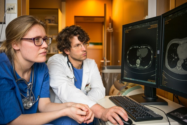
Teleradiology – perfect solution
As it was mentioned, the description of X-ray images and other images from radiological examination should be always performed by a specialist. However less and less physicians decide on this specialty. Therefore, teleradiology is the solution for aproblem with limited access to qualified specialists. It allows to contact high class radiologists, who describe the radiological images from any place in Poland or the world. Thank to this even small medical facilities can afford it and provide high quality services to their patients.